Why Evolution is True is a blog written by Jerry Coyne, centered on evolution and biology but also dealing with diverse topics like politics, culture, and cats.
Well, this is the last batch of submitted photos, but I hope for me. Don’t dash my hope!
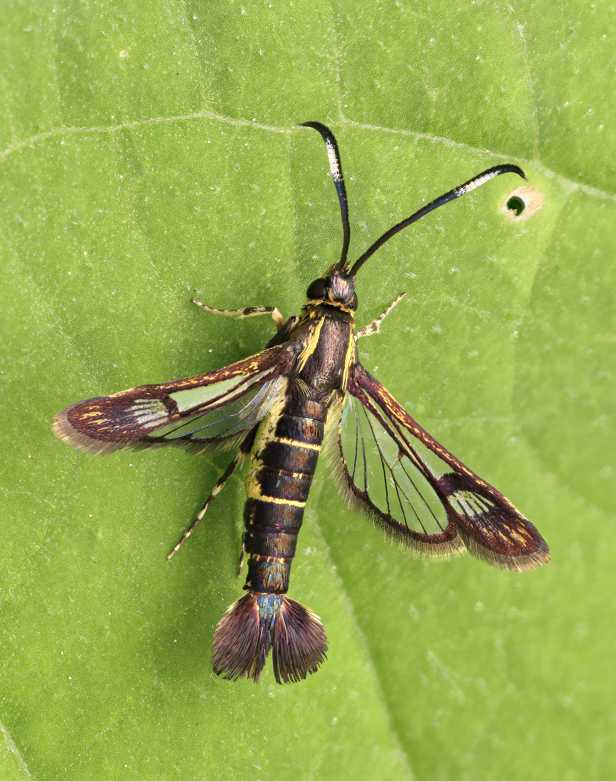
Today we have a lovely text-and-photo post by Athayde Tonhasca Júnior, featuring a bizarre and mimetic beetle. Athayde’s captions are indented, and you can enlarge the photos by clicking on them.
Fabulous pretenders
Termites, cockroaches’ sophisticated cousins (Order Blattodea), live in intricate, organized societies with division of labour and a caste system. The mound-building species are also skilled engineers, constructing temperature-controlled, ventilated nests that protect their inhabitants from the harsh conditions of the outside world. Colonies may comprise millions of individuals, including eggs, larvae and workers. Just like other social insects, termites have to be on guard against many an envious enemy: their cosy nests are tempting to would-be squatters, with the even more tempting bonus of being packed with energy-rich morsels.
Damage to a nest of Formosan subterranean termites brings hoards of workers and soldiers with dark, oval shaped heads scrambling to repair the hole. Termites shown about 4 times actual size. USDA photo by Scott Bauer.
Termites are mostly successful in keeping invaders at bay, but a sizable group of outsiders has evolved skills that allow them to breach those defences. These are the termitophiles: macro-organisms that live in association with termite colonies. Termitophiles, ranging from harmless inquilines to predators and parasites, rely on chemical mimicry and numerous morphological and behavioural adaptations to avoid detection and mingle with their hosts.
Among the many impostors, rove beetles from the subfamily Aleocharinae are particularly noteworthy. This is a huge group (~16,000 species) within the humongous Staphylinidae family, which comprises some 66,000 species, one of the largest families of organisms. Many aleocharines are myrmecophilous (associated with ants); some 670 species are termitophilous.
Aleocharines have reached extraordinary levels of deception, but two termitophilous species of the genus Austrospirachtha from northern Australia – the only known species so far – take their art to a new level. On first seeing their images, one may think they are AI-generated. Or pranks devised by putting together bits of different insects, entomological versions of the Piltdown Man hoax.
The termite puppets on their backs, complete with dangling pseudo-appendages that resemble antennae and legs, fool their hosts into accepting them as nestmates. You may see these beetles as rough simulacrums of the real thing, but in the pitch-dark confines of a termite nest, mimicry is based on palpation rather than vision (Watson, 1973). The mouthparts of A. carrijoi are very small, which suggest it dupes termite workers to feed it, a process known as trophallaxis (Zilberman & Pires Silva, 2023). Presumably, the same happens with A. mimetes.
We know very little about these beetles or any other symbiotic aleocharines. But the rare insights into their outlandish appearances are glimpses of the marvellous workings of natural selection.
References
Pires Silva, C.M. 2024. Cladistic analysis, taxonomic revision & biological notes of the termitophilous genus Xenogaster Wasmann, 1891 (Staphylinidae, Aleocharinae, Corotocini). Master’s Dissertation, Universidade de São Paulo, Brazil.
Watson, J.A.L. 1973. Austrospirachtha mimetes, a new termitophilous corotocine from Northern Australia (Coleoptera: Staphylinidae). Journal of the Australian Entomological Society 12: 307-310.
Zilberman, B. et al. 2019. Viviparity in Staphylinidae and reproductive behavior of Corotoca Schiødte, 1853. Papéis Avulsos de Zoologia 59: e20195919.
Zilberman, B. & Pires Silva, C.M. 2023. A new species and morphological notes on the remarkable termitophilous genus Austrospirachtha Watson from Australia (Coleoptera: Staphylinidae: Aleocharinae). Zootaxa. 5336: 424-432.
Regular Mark Sturtevant (as opposed to Irregular Mark Sturtevant) has sent in some lovely insect photos to fill the nearly-empty reserve of pictures. Please send any good wildlife photos you have. Thanks!
Mark’s captions are indented, and you can enlarge his photos by clicking on them. Be sure to check out the female Tussock Moth caterpillar with vestigial wings (it’s the eighth photo).
Here is another dispatch of local insects and spiders, all photographed either in area parks near where I live in eastern Michigan, or as staged shots at home. Let’s start with a couple of dragonflies. First up is a Green Darner (Anax junius), which ranks pretty large among the dragonflies found here. As is almost always the case, this individual is a female, since they perch frequently and are much easier to approach than males, which I only rarely manage to photograph. Do you see the tiny yellow critter on the dragonfly? That is a Globular Springtail, and it must have walked or jumped onto the dragonfly from nearby foliage. There may be a few more of them in the picture as well, though I’m not certain. Springtails are tiny soil arthropods, and they are extremely abundant. When seen up close, Globular Springtails are arguably adorable, as you can see in the linked image. I’ve searched for them for some time but have only rarely encountered them, despite claims that they are exceedingly common. Perhaps our local species spends more time up on foliage rather than in the soil, meaning I’ve been looking in the wrong places!:
The next dragonfly is a male Skillet Clubtail (Gomphurus ventricosus), named for the large, pan-like club on the end of its abdomen. I have to drive a few hours south to a particular park to see them, and of course this one is perching on poison ivy which covers much of the area there. That park hosts 8 or 9 species of clubtails according to a dragonfly documentation site called Odonata Central. I’ve photographed most of those species with only two left to find:
A field near my workplace has a ridiculously high population of Chinese Praying Mantids (Tenodera sinensis). I suspect this is the lingering result of someone having released a large number of egg cases several years ago, combined with a robust population of grasshopper prey. Shown here is a young June nymph. I can return later in the summer to find several large adults in no time at all:
Next up are some lepidopterans. First is a Hackberry Emperor butterfly (Asterocampa celtis). They are often common along forest margins where their host Hackberry trees [Celtis occidentalis] are concentrated. Their caterpillars, however, seem to be quite secretive, as I’ve seen only a few of them:
Next is an inchworm caterpillar, though I don’t have an identification for it. Do you see the tiny mites?:
The insect shown next is a moth, although it makes a very convincing wasp mimic. This is the Eupatorium Borer Moth (Carmenta bassiformis). The larvae feed on the roots of Ironweed and Joe Pye Weed, both of which are highly valued native wildflowers because they are very much favored by butterflies:
The flamboyant caterpillar in the next image is a White-marked Tussock Caterpillar (Orgyia leucostigma). I’ve tried several times to get an acceptable focus-stacked image of one of these. What makes them difficult is the combination of all those oddly angled sticky-out bits, which challenge the shallow depth of field inherent in this kind of photography, along with the fact that these caterpillars absolutely will Not Hold Still. As a result, the stacking is done on the fly, followed later by long hours of manual reassembly of the jumbled focus stack. This image is by far my most successful attempt:
I kept this caterpillar in hopes that the moth emerging from the cocoon would be a female. Why? Well—check out what came out! It was a female! Females have barely visible vestigial wings, and I had never seen one before. She will sit here, unmoving, until a male finds her. This strategy saves a great deal of energy that can instead be used for laying eggs. The winged males are unremarkable, and while I’ve probably seen them, I don’t think I’ve ever photographed one:
Finally, here are three images of jumping spiders I’ve been wanting to share together for quite some time. The Dimorphic Jumping Spider (Maevia inclemens) is a species I regularly see around the outside of our house. The name refers to the males having two distinct color morphs. The images below are manual focus stacks, photographed in staged settings on the ‘ol dining room table. First is a female with long-legged fly prey, which conveniently provides a sense of scale since the fly is about the size of a mosquito:
Next are the two male color morphs. The first somewhat resembles the females, while the other is very different and comes with distinctive hair decorations. This is an older image, but I’m bringing it back so all three forms can finally be shown together. It’s surprising that they are all the same species!
I recently learned that the two male forms use different courtship displays for females, yet are considered equally successful in the mating game. I’m sure Jerry can steer things in the right direction if I am in error here, but having different male forms with different mating strategies does not seem unusual in the animal kingdom. Examples I’m familiar with include lizards, crustaceans, and fish, although in those cases the different males include those that are aggressive and territorial, and others that win by being sneaky. But here, I don’t see how either male is territorial, and apparently both have courtship displays for females.
One of my goals for next season will be to try to document their different courtship behaviors. That seems do-able, since I can expect to find several of them again next summer.
Today we have a photo-and-text submission from Athayde Tonhasca Júnior on fly migration. It’s a subject dear to my heart as I used to work on it, publishing three papers on migration in Drosophila. Athayde’s subject, though, is hoverflies, not fruit flies. His captions are indented, and you can enlarge his photos by clicking on them. Note: I changed Athayde’s words “hover flies” to the more common usage “hoverflies,” but Athayde notes that most entomologists use the two-word rather than one-word description.
On the road again, goin’ places that I’ve never been
Sometime between 1400 and 1200 BC, Yahweh (aka God) decided it was time to nudge the Egyptians to let their captive Israelites go. Yahweh could have tried diplomacy, but in his infinite wisdom he concluded that “The Egyptians shall know that I am the LORD”. And there was no better way to let the Pharaoh and his people know who the bigwig was around there than by punishing them with a series of plagues. Of the ten celestial disasters inflicted upon the Egyptians, two involved mosquitoes (or midges) and flies, which probably were also the agents behind another two plagues manifested as infectious diseases of people and livestock. Yahweh understood very well the efficacy of some flies (order Diptera) and pathogens to wreck revenge – after all, he created them.
The tales of pestilent flies depicted in the book of Exodus could have been inspired by real events, as pest infestations and epidemics were recurrent in the ancient world. Fly outbreaks are facilitated by these insects’ capability to disperse for long distances and arrive at new locations suddenly and in massive numbers. There are no better examples of these efficient colonisers than hoverflies or syrphid flies (family Syrphidae) such as the marmalade (Episyrphus balteatus) and the migrant (Eupeodes corollae) hoverflies. Each autumn, they leave Britain and head south to spend the winter in southern Europe and the Mediterranean. Their offspring move northwards in the spring, lay eggs, and the new generation sets out on the cycle again. Researchers have estimated that up to four billion marmalade and migrant hoverflies cross the English Channel to and from Great Britain every year. This represents 80 tons of biomass. If you are impressed by these figures, you should know that hoverflies account for a fraction of insects’ latitudinal migrations known as ‘bioflows’: about 3.5 trillion insects, or 3200 tons of biomass, migrate into southern Britain annually (Wotton et al., 2019). Insect bioflows pour vast amounts of nutrients (particularly nitrogen and phosphorus) and countless prey, predators, parasites and herbivores into ecosystems, but we have only a vague understanding of their impact on food webs and local species.
These hardy wanderers have another particularity of significant ecological importance: they transport pollen grains.
Most flies have no pollen-collecting structures and have few ‘hairs’ (setae), which are important pollen gatherers. These are negative marks for candidates to the pollinators’ club, but some flies compensate their shortcomings by their massive numbers. Each marmalade and migrant hoverfly carries an average of 10 pollen grains from up to three plant species on their journey into Britain. That’s paltry compared to a bee, but altogether, those flies bring in 3 to 8 billion pollen grains on each inward journey.
Pollen importation via flies is a recurrent phenomenon. In Cyprus, warm temperatures and favourable winds bring millions of insect migrants from the Middle East region, more than 100 km to the east. Flies make up nearly 90% of these bioflows, and many of them are loaded with pollen (Hawkes et al., 2022).
Pollen-loaded flies can turn up anywhere the wind takes them, even to specks of dry ground in the middle of nowhere. Over a two-month period, 121 marmaladehover flies reached a North Sea oil rig approximately 200 km from Aberdeen, UK. Over 90% of these flies had pollen attached to them, sometimes from eight plant species. Based on pollen barcoding and wind trajectory modelling, it was estimated that these flies traversed from 265 to 500 km of open water in a single journey, probably from the Netherlands, Germany and Denmark (Doyle et al., 2025).
Flies’ long-distance pollen transfers may help connect isolated plant populations, such as in fragmented habitats, but we don’t know much about the ecological implications. However we do know that their contribution can be important. In continental Europe, wild carrot (Daucus carota) depends on a range of insects for pollinators, especially bees. But bees are absent from La Foradada, a 1,6 ha Mediterranean islet about 50 km off the Spanish coast. In this solitary spot of land, D. carota subsp. commutatus relies on the accidental arrival of common drone flies for its pollination (Pérez-Bañón et al., 2007).
Butterflies, bumble bees, moths and dragonflies are known travellers, but we know much less about migrant flies, which may have significant roles in pollination ecology. We just have to pay more attention to these unpretentious pilgrims.
References
Doyle, T.D. et al. 2025. Long-range pollen transport across the North Sea: Insights from migratory hoverflies landing on a remote oil rig. Journal of Animal Ecology 94: 2267–2281.
Hawkes, W.S.L. et al. 2022. Huge spring migrations of insects from the Middle East to Europe: quantifying the migratory assemblage and ecosystem services. Ecography e06288.
Pérez-Bañón, C. et al., 2007. Pollination in small islands by occasional visitors: The case of Daucus carota subsp. commutatus (Apiaceae) in the Columbretes archipelago, Spain. Plant Ecology 192: 133-151.
Wotton, K.R. et al. 2019. Mass seasonal migrations of hoverflies provide extensive pollination and crop protection services. Current Biology 29: 2167–2173.
Send ’em in, please, or we’ll be empty over the holiday season.
Today’s photos of butterflies come from Pratyaydipta Rudra, a statistics professor at Oklahoma State University. Pratyay’s captions and IDs are indented, and you can enlarge the photos by clicking on them. Pratyay and his wife Sreemala have a big bird-and-butterfly website called Wingmates.
Here are various other butterflies photographed in Oklahoma, most of them at our backyard.
Gulf Fritillary (Dione vanillae) eyeing a flower to land on, but it was already occupied by a skipper:
Common Buckeye (Junonia coenia) on cosmos. These plants are hardy and attract many pollinators:
Another commoner in our area – American Lady (Vanessa virginiensis):
Painted Lady (Vanessa cardui), looks very similar to the American Lady, but one can spot the difference if they look carefully. They have the widest distribution of any butterfly, found on all continents except Antarctica and Australia:
Another Painted Lady (Vanessa cardui) on our cosmos. Mid-day harsh light is terrible for bird photography, but it can be worked around when photographing butterflies. In fact, I quite like the glow that comes from the sun hitting the butterflies from directly above:
Closeup of a Painted Lady (Vanessa cardui). They have a long “nose” (actually, elongated mouthpart), but can’t beat the American Snout (Libytheana carinenta) in this regard:
The next two images are for the readers to guess which one is an American Lady and which is a Painted Lady. Can you tell what this one is?:
Viceroys (Limenitis archippus) look similar to Monarch, but they are smaller and have a black line across the hindwing that the Monarch doesn’t. They were thought to be a Batesian mimic of Monarch for a long time, but are now considered a Mullerian mimic where both species are unappealing to predators and evolve similar appearances, reinforcing predators’ avoidance of either one:
Not all butterflies visit flowers. This Question Mark butterfly (Polygonia interrogationis), for example, only visits flowers when its usual food sources – rotting fruit, plant sap, dung etc., are not easily available:
Tawny Emperors (Asterocampa clyton) have a similar diet:
Hackberry Emperor (Asterocampa celtis), another similar butterfly from the same family. They are quite common in our area:
Many animals are venomous, but in most cases the exact proteins involved in causing pain or death are unknown, and even in those cases the genes producing them have not been identified, counted or mapped. If you’re interested in the evolution of venom, what its precursors are, and how venomous animals avoid poisoning themselves, you have to know this kind of stuff.
A new paper in Proc. Nat. Acad. Sciences (click screenshot below to read for free, or find the pdf here) answered several of these questions in the venomous caterpillar of the mottled cup moth (Doratifera vulnerans), shown below. It’s from Australia, and is described in Wikipedia this way:
It is known for its caterpillar having unique stinging spines or hairs that contain toxins, for which the scientific name is given that means “bearer of gifts of wounds”. Chemical and genetic analysis in 2021 show that its caterpillar contains 151 toxins, some of which have medicinal properties
That earlier paper, from 2021 and including some of the same authors as the one we discuss today, did indeed identify 151 proteins (peptide are bits of proteins or short chains of amino acids) that were in the toxins, but did not know which genes produced them, how the genes were arranged, what the closest relatives of the genes were, and how many of the 151 “toxins” were really toxic (the word “toxin” there and in the present paper do not mean that the substances were toxic, but that they were simply a component of the extracted toxins). However, the authors, some on the paper I’m highlighting today, did identify two genuine toxins that caused pain: the peptides Dv12 and Dv11.
Look at this thing! It’s clearly aposematic, meaning that it has bright warning coloration that predators can recognize and learn to avoid. And you can see those nasty spines. In the earlier paper they extracted toxins from related species and tested them by injecting them into mice tails, guinea pigs, and human volunteers. That earlier paper also adds this about the species name:
This species, whose binomial name etymologically means “bearer of painful gifts,” is a common culprit of caterpillar envenomations in Australia.
That means that many Aussies get stung by these things, probably inadvertently. Would you touch an animal that looks like this?:
On to the new paper, and I’ll try to be brief as it’s long and complicated.
1.) First, the authors sequenced the entire caterpillar genome (remember, it’s the same as the adult moth genome).
2.) Then, knowing the sequences of the proteins known from previous work on toxins, they could find the genes producing them by matching the protein sequence to the DNA sequence that could produce these proteins. Of the 151 proteins in caterpillar venom known from the prvious work, they mapped 149 of them to 115 sites in the genome
3.) Of the 115 sites, 35 were products of single genes, while 80 (70%) of the total, were members of gene families consisting of two or more similar genes (sometimes many genes) with similar sequences. Here’s a map of the “toxin gene” locations on the insect’s 13 chromosomes. The blue dots are the genes existing in single copies, orange dots are clusters of genes previously grouped together by protein-sequence similarity, and pink dots are genes that were newly identified, surely as part of gene families, in the present study. This conclusion comes from their sequence similarity and they physical grouping on two chromosomes.(The size of the dots indicates the number of genes that are part of a contiguous group. Click to enlarge:
So we know that genes found in venom are very often the product of gene duplications, either of single genes becoming two (this can happen via unequal crossing-over during meiosis or by other methods), producing two initially identical genes side by side or whole groups of them (“tandem duplications”). Once a gene has been duplicated, the original copy can then keep its original function, while the other copies, not being “needed,” are free to evolve other functions. Many genes we’re familiar with, like our own globins and immunoglobulins, evolved by gene duplication followed by divergence of the duplicated copies.
Where did the genes making venom proteins come from? This is the key evolutionary question answered here and, to some extent, in the previous paper. They evolved from ancestral genes in the moth’s immune system that evolved to attack microbes, the so-called “antimicrobial peptides” (AMPs), also known as cecropins. The ancestral AMP proteins, nearly identical to their original form and function, kill bacteria (prokaryotes) by disrupting the bacterial membranes. Insects still need to kill microbes!
Clearly, the proteins in venom have evolved by natural selection modifying ancestral genes used to kill bacteria. Now they are used to repel predators. Natural selection causing this divergence was implicated by looking at sequence differences, as there are ways of showing what sequence differences evolve faster than expected under either the slower processes of genetic drift or “purifying” selection that conserves structure. They found that most of the venom-adapted proteins that evolved from cecropins did evolve under natural selection, while the descendants of cecropins that retained their original anti-microbial proteins were under purifying selection to retain their sequence. It’s clear, then, that the insect still needs genes to attack bacteria. It’s just that some of them have been repurposed, often through gene duplication and divergence, to repel predators. (The authors have a way of assessing “pain” by measuring the increase in calcium concentration in cells grown in vitro and exposed to venom. This happens when the two investigated proteins are used.)
Here is a complicated family tree of cecropin genes in black used to kill microbes. The genes found in venom are in the red box (“venom adapted”). You can see that they are related to cecropin genes but branched off fairly recently (probably four or five million years ago). The venom genes are in the red box that I’ve added, and their relationship as being derived from ancestral AMP genes is very clear. (The “canonical” genes in green are antimicrobial proteins closely related in sequence to the venom genes.
So, now we know where the genes in venom come from. What we do not know is how many of those genes are essential in venom, either causing pain or doing other stuff that venom needs to do. At least two of them cause pain, but there are probably more, for they haven’t all been tested. And some of the other genes are probably involved in dismantling cell walls in potential predators. The authors tested several of the venom proteins and also found that, as in their AMP ancestors, they disrupt cell covering, in this case eukaryotic cell membranes.
Finally, the big question: If the caterpillar makes venom, why doesn’t it poison itself? Here’s how the authors answer that question (I’ve put the answer for this species is in bold).
Animals that produce toxins, either for innate immunity or as venom toxins, must employ strategies to protect themselves from toxicity. Such protective mechanisms include production of toxin inhibitors, storage in inactive form, mutations in their own ion channels that confer resistance, alteration of lipid bilayer compositions, and compartmentalization of toxins separate from body tissues. In the case of limacodid venom peptides, the venom is compartmentalized into the cuticle-lined venom reservoir inside venom spines, preventing the toxin from coming into contact with cells other than the secretory cells that produce them. Thus, compared to canonical cecropins, venom-adapted cecropins may also be released from pressure to avoid activity against animal cells.
There are other findings in the paper that will be of interest primarily to those studying genomic evolution. For example, many of the venom proteins still retain some weak antimicrobial activity, so the idea that genes completely lose their ancestral function when they gain a new one doesn’t hold in this case.
Below you can see the adult moth because, remember, they studied caterpillar venoms, and many of those genes are probably turned off in the adult. But adult and caterpillar carry the exact same genes, of course; their different bodies, physiology, and behavior rest on the differential turning on and off of these genes at different life stages. And that remains a big mystery: how do such different life stages evolve, with each step of the evolution being adaptive?
From The Australian Museum, photo credits at bottom (click to enlarge), image by Lyn Craggs.
Today I’m putting up all the singletons and smaller batches sent to me. Readers’ captions and IDs are indented.
From Allen Jones (I don’t know the species). It’s in the UK; can readers help?
This was at a local bird of prey centre near Edinburgh. The eagle’s perched on me. The owner said that this boy or girl (not sure) would soon be the oldest eagle that had ever lived in the UK. He said it was around 50 years old.
From A. C. Harper:
Ordinary urban pigeons huddling together for warmth… but they are on a support of a bridge crossing an arm of the river Soar at Leicester. This was once open fields, then industry including extensive railway sidings, a foundry, a stone masons, several mills (afer canalization of this part of the river) and wharves for unloading coal. As you can see the industrial areas have now gone and are being replaced with houses and apartments. But the pigeons linger on ready for my cellphone, a Pixel 10 pro.
From Susan Harrison, who promises that there is more to come.
Thought you’d enjoy seeing this big Belizean kitty! He was lounging in the road last night as we drove into the Rio Bravo Conservation Area. He could not have cared less about us, which is always a nice thing to experience with wildlife. Anyway, this is just a teaser, and I’ll send a batch for RWP soon.
From Bryan Lepore.
These little Eastern cottontails [Sylvilagus floridanus] are in mid-ish Massachusetts outside of Framingham, and in my back yard. First photo June 26, second June 29:
From Nicole in Pennsylvania: A wild cottontail that comes to humans for apple slices. Her name is Petunia:
Convergent evolution, pictures by Martin Riddle.
The Hawk Moths, aka Hummingbird Moths [JAC: Family Spingidae] love the nectar in resident maintained gardens at Brooksby Village Peabody, Massachusetts:


